
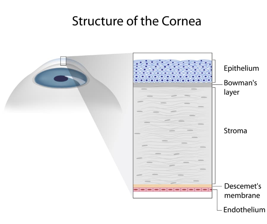
The cornea can be referred to as, “the window to the eye”. It is in the front of the eye and covers the iris, pupil and anterior chamber. The human cornea is comprised of six different cell layers: Epithelium, Bowman’s Layer, Stroma, Dua’s Layer, Descemet’s Membrane and Endothelium.
The epithelium is the outermost layer of the cornea and accounts for about 10% of the cornea tissue’s thickness. The epithelial layer has two main functions: to obstruct the passage of foreign materials and to absorb oxygen and nutrients from the eye’s tears to provide a smooth surface layer. The nutrients that are absorbed from the tear layer are distributed to the remainder of the cornea via the epithelium. The Corneal epithelium is supplied by thousands of nerve endings making the cornea the most sensitive area on the body (resulting in significant discomfort when the epithelial cell layer is disturbed as a result of trauma or disease).
Bowman’s layer consists of a transparent layer of tissue and exists directly under the epithelial cell layer. This tissue is composed of strong layered protein fibers referred to as collagen. If injury occurs at the Bowman’s layer, scars can result during the healing process. These scars can result in some vision loss, especially if they are large in nature or located centrally.
The third cell layer of the cornea is called the, stroma. This cell layer accounts for nearly 90% of the cornea’s thickness and is composed primarily of water and collagen. The collagen provides the cornea with strength, flexibility and structural integrity. This collagen layer is most important in generating the cornea’s light-conducting transparency, which in turn, provides the eye with the majority of its focusing power.
Dua’s layer is the thinnest layer of the cornea, measuring only 15 microns – less than half the size of a human hair! This portion of the cornea is new to the scientific world. In fact, it was identified in the summer of 2013 by researcher, Harminder Dua of the UK. Due to its recent discovery, not much is known about the Dua’s layer. Ophthalmologists and scientists are hopeful that this discovery will help make corneal procedures safer and simpler for patients who have damage to this portion of the eye.
The Descemet’s membrane exists beneath the stroma and is a thin, yet strong tissue layer. The main objective of the Descemet’s membrane is to act as a barrier against infection and injuries to the eye. Again, the Descemet’s membrane is comprised primarily of collagen fibers, but the fibers are different than those present in the stromal layer. In fact, the collagen fibers present in the Descemet’s membrane are created by the endothelial layer that exists below this membrane. Fortunately, this allows for the Descemet’s membrane to be restored readily if injury occurs.
Finally, the innermost layer of the cornea is the endothelium, which is an extremely thin cell layer. This thin endothelial cell layer plays a crucial role in maintaining a clear cornea. In a normally functioning eye, fluid drains slowly from the inside of the eye into the stroma (the middle corneal layer). The endothelium acts to pump the excess fluid out of the stroma. If the endothelial layer becomes ineffective at pumping out this excess fluid, the stroma begins to swell with water. If this occurs, it can lead to the stromal layer becoming swollen, hazy and eventually opaque causing blurred vision.
In a healthy cornea, the endothelium properly balances the amount of fluid moving into the cornea and the amount of excess fluid that is pumped out of the stroma; however, if the endothelial cells are destroyed by trauma or disease, they are lost forever. Endothelial cells are not regenerated, so if too many of the cells are damaged, the cornea becomes swollen and blurred vision or blindness can result. The most common means of damaging the endothelial layer is through aging, inherited eye diseases (such as Fuchs’ Corneal Dystrophy), ocular trauma, and previous intraocular surgeries. Mild cases of corneal swelling can be improved by use of topical drops and ointments (such as Muro 128 2% drops and Muro 128 5% ointment). More severe cases of corneal edema require corneal transplantation to replace the endothelial layer, reviving some of the cornea’s ability to focus light rays.